Male mating (WP2287)
Caenorhabditis elegans

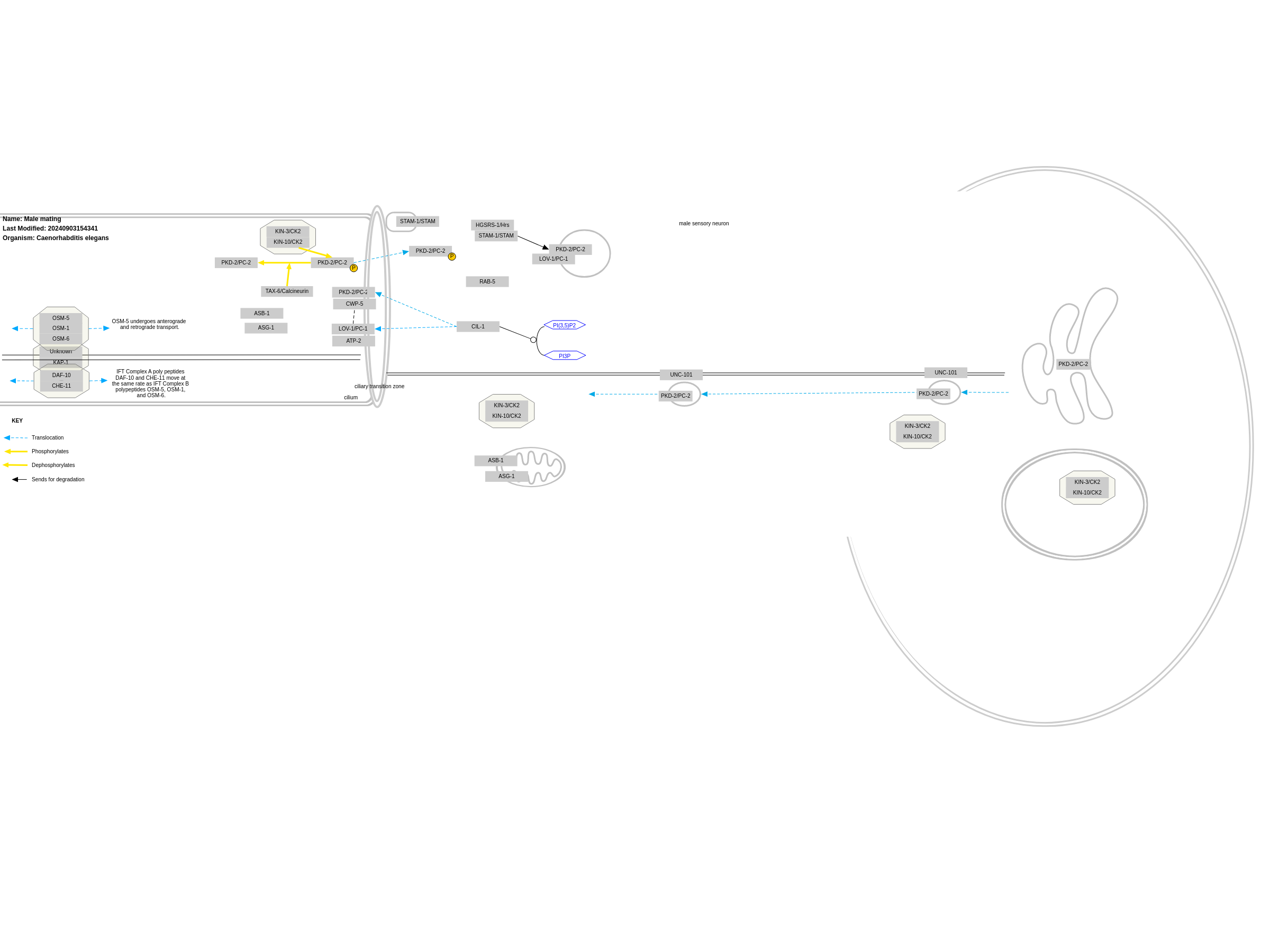
Caenorhabditis elegans males exhibit sterotypic and invariant mating behavior starting the moment the animal senses a hermaphrodite and ending with insemination. This complex behavior has been broken down into six steps or sub behaviors: male response to hermaphrodite contact, backwards movement along her body, sharply turning in a ventral coil upon reaching the head or tail, continued backing until his tail contacts the vulva (vulval location), spicule insertion, and ejaculation into the hermaphrodite uterus. Each of these sub behaviors have been molecularly dissected and it has been found that two of these sub behaviors, male response to hermaphrodite contact and vulval location involve similar molecules. In particular these behaviors involve cell autonomous signaling through LOV-1 and PKD-2, which are homologs of human polycystin kidney disease (PKD) associated genes PC-1 and PC-2 respectively. Studies in C. elegans have shown that these genes likely have a sensory function rather than structural or development role in the cilia of male sensory neurons. The identification and characterization of LOV-1 and PKD-2 in C. elegans has lead to insights and new avenues of inquiry in the study of human PKD polycystin pathways.
For a description of pathway objects, see the WikiPathways Legend.
Authors
Karen Yook , Stefan Raats , Finterly Hu , and Egon WillighagenActivity

Discuss this pathway
Check for ongoing discussions or start your own.
Cited In
Are you planning to include this pathway in your next publication? See How to Cite and add a link here to your paper once it's online.
Organisms
Caenorhabditis elegansCommunities
WormBaseAnnotations
Pathway Ontology
protein sorting pathway lysosomes based pathway of protein degradationDisease Ontology
autosomal dominant polycystic kidney diseaseCell Type Ontology
ciliated cell| Label | Type | Compact URI | Comment |
|---|---|---|---|
| PKD-2/PC-2 | GeneProduct | wormbase:Y73F8A.1 | PKD-2 phosphorylation state appears to modulate its function and ciliary localization, with S534A and S534D reflecting two extreme states.CK2-phosphorylated PKD-2 is dephosphorylated by calcineurin. |
| LOV-1/PC-1 | GeneProduct | ensembl:WBGene00003058 | LOV-1 localizes to intracellular membranes |
| PKD-2/PC-2 | GeneProduct | wormbase:Y73F8A.1 | PKD-2 localizes to intracellular membranesFunctional localization of PKD-2 is in the ciliary plasma membrane and in intracellular membranePKD-2 degradation does not require K48 polyubiquitination and is likely due to STAM-1-Hrs-mediated degradation. |
| UNC-101 | GeneProduct | ensembl:WBGene00006829 | UNC-101 acts at a somatodendritic sorting step to restrict PKFD-2, along with other ciliary receptors, to the dendritic compartment. |
| KIN-3/CK2 | GeneProduct | ensembl:WBGene00002191 | kin-3 and kin-10 are coexpressed with lov-1 and pkd-2 in the male-specific CEM head neurons and ray RnB and hook HOB tail neuronsKIN-3::GFP and KIN-10 GFP are enriched in cilia and also found in cell bodies (including nuclei), dendrites, and axons. |
| STAM-1/STAM | GeneProduct | ensembl:WBGene00004109 | STAM-1A specifically associates with the LOV-1 C-tailstam-1 and pkd-2 are clearly coexpressed in male-specific ciliated CEM, ray B-type(RnB), and hook HOB sensory neuronsSTAM-1A::GFP labels puncta and accumulates at ciliary bases, but is obviously excluded from the cilium proper of CEM, RnB and HOB neuronsSTAM-1A co-localizes at the ciliary base with LOV-1 and PKD-2STAM-1 and HGRS-1 expression completely overlaps in cell bodies and ciliary bases of polysystin-expressing neuronsSTAM-1A. but not STAM-1B, specifically acts in the polycystin down-regulation process.STAM-1A binds, sorts, and targets the polycystin complex for lysosomal degradation |
| RAB-5 | GeneProduct | ensembl:WBGene00004268 | STAM-1 and RAB-5 collocate in the cell bodies and ciliary bases of polycystin-expressing neurons. |
| HGSRS-1/Hrs | GeneProduct | ensembl:WBGene00004101 | STAM-1 and HGRS-1 expression completely overlaps in cell bodies and ciliary bases of polysystin-expressing neurons. |
| PKD-2/PC-2 | GeneProduct | wormbase:Y73F8A.1 | PKD-2 is synthesized in the ER and packaged into vesicles that are transported to the ciliary base and inserted onto the ciliary membrane. |
| CIL-1 | GeneProduct | ensembl:WBGene00086546 | |
| ASB-1 | GeneProduct | ensembl:WBGene00000206 | ASB-1 and ASG-2 localize to male-specific sensory cilia as well as mitochondria. |
| OSM-1 | GeneProduct | ensembl:WBGene00003883 | |
| DAF-10 | GeneProduct | ensembl:WBGene00000906 | |
| OSM-5 | GeneProduct | ensembl:WBGene00003885 | OSM-5 localizes to cilia through its TPR repeats through intraflagellar transport |
| LOV-1/PC-1 | GeneProduct | ensembl:WBGene00003058 | LOV-1 localized to cilia by CIL-1LOV-1 is expressed in male specific sensory neurons...with basodendritic subcellular localization in cell body and sensory endingslov-1 is exclusively expressed in three categories of adult male sensory neurons: the rays, the hook, and the head CEMslov-1 and pkd-2 are expressed in male-specific sensory neurons and are localized to the similar region of the same cell.lov-1 and atp-2 are coexpressed in the tail ray B neurons and HOB hook neuron...as well as the male-specific CEM head neurons. |
| CHE-11 | GeneProduct | ensembl:WBGene00000490 | |
| OSM-6 | GeneProduct | ensembl:WBGene00003886 | |
| KAP-1 | GeneProduct | ensembl:WBGene00002182 | IFT Complex A poly peptides , DAF-10 and CHE-11 move and that motility is at the same rate as IFT Complex B polypeptides, OSM-1, OSM-5 and OSM-6, and KAP (a subunit of the kinesin motor). |
| CWP-5 | GeneProduct | wormbase:F48C11.2 | |
| PKD-2/PC-2 | GeneProduct | wormbase:Y73F8A.1 | PKD-2 localized to cilia by CIL-1PKD-2 GFP accumulates in a ring around the ciliary transition zones.Functional localization of PKD-2 is in the ciliary plasma membrane and in intracellular membranePKD-2 protein is hghly enriched in sensory cilia that are directly exposed to the environmentWithin the ciliary region, PKD-2::GFP is distributed throughout the cilium, but primarily concentrated at the ciliary base, which corresponds to the distal-most dendrite and transition zone regions.In wild-type adult males, PKD-2 is distributed along the ciliary axoneme and accumulates in the transition zone between the cilium and dendrite. |
| ATP-2 | GeneProduct | ensembl:WBGene00000229 | ATP-2 is identified as one candidate that physically interacts with the PLAT domain of LOV-1ATP-2 and other ATP synthase components were found to localize to the cilia of male-specific sensory neurons.ATP-2::GFP clearly colocalizes with PKD-2::DsRed2 in CEM cilia.ATP-2 physically interacts with LOV-1 |
| ASG-1 | GeneProduct | ensembl:WBGene00000209 | ASB-1 and ASG-2 localize to male-specific sensory cilia as well as mitochondria. |
| KIN-10/CK2 | GeneProduct | ensembl:WBGene00002196 | kin-3 and kin-10 are coexpressed with lov-1 and pkd-2 in the male-specific CEM head neurons and ray RnB and hook HOB tail neuronsKIN-3::GFP and KIN-10 GFP are enriched in cilia and also found in cell bodies (including nuclei), dendrites, and axons.CK2-phosphorylated PKD-2 is dephosphorylated by calcineurinCK2, lov-1, and pkd-2 act in the same genetic pathway regulating mating behavior.CK2 modulates PKD-2 function.PKD-2(S534) is one but not the only in vitro CK2 phosphorylation site. |
| TAX-6/Calcineurin | GeneProduct | ensembl:WBGene00006527 | TAX-6::GFP also localizes to cilia of these polycystin-expressing neuronsCK and calcineurin/protein phosphatase 2B(PP2B) modulate PKD-2 function and ciliary localizationCK2 and calcineurin function antagonistically to regulate PKD-2 phosphorylation stateCK2 phosphorylated PKD-2 is dephosphorylated by calcineurinIn the male, we observed tax-6 expression in the CEM, HOB, and ray RnB neurons, with expression noticeably absent from ray 6, which is very similar to lov-1 and pkd-2 expression patterns |
| STAM-1/STAM | GeneProduct | ensembl:WBGene00004109 | GFP-tagged STAM-1 fusion protein localized to cytoplasmic and dendritic puncta resembling endosomes |
| KIN-10/CK2 | GeneProduct | ensembl:WBGene00002196 | kin-3 and kin-10 are coexpressed with lov-1 and pkd-2 in the male-specific CEM head neurons and ray RnB and hook HOB tail neuronsKIN-3::GFP and KIN-10 GFP are enriched in cilia and also found in cell bodies (including nuclei), dendrites, and axons. |
References
- A polycystic kidney-disease gene homologue required for male mating behaviour in C. elegans. Barr MM, Sternberg PW. Nature. 1999 Sep 23;401(6751):386–9. PubMed Europe PMC Scholia
- An autosomal recessive polycystic kidney disease gene homolog is involved in intraflagellar transport in C. elegans ciliated sensory neurons. Qin H, Rosenbaum JL, Barr MM. Curr Biol. 2001 Mar 20;11(6):457–61. PubMed Europe PMC Scholia
- The Caenorhabditis elegans autosomal dominant polycystic kidney disease gene homologs lov-1 and pkd-2 act in the same pathway. Barr MM, DeModena J, Braun D, Nguyen CQ, Hall DH, Sternberg PW. Curr Biol. 2001 Sep 4;11(17):1341–6. PubMed Europe PMC Scholia
- ATP-2 interacts with the PLAT domain of LOV-1 and is involved in Caenorhabditis elegans polycystin signaling. Hu J, Barr MM. Mol Biol Cell. 2005 Feb;16(2):458–69. PubMed Europe PMC Scholia
- Mating worms and the cystic kidney: Caenorhabditis elegans as a model for renal disease. Lipton J. Pediatr Nephrol. 2005 Nov;20(11):1531–6. PubMed Europe PMC Scholia
- Casein kinase II and calcineurin modulate TRPP function and ciliary localization. Hu J, Bae YK, Knobel KM, Barr MM. Mol Biol Cell. 2006 May;17(5):2200–11. PubMed Europe PMC Scholia
- General and cell-type specific mechanisms target TRPP2/PKD-2 to cilia. Bae YK, Qin H, Knobel KM, Hu J, Rosenbaum JL, Barr MM. Development. 2006 Oct;133(19):3859–70. PubMed Europe PMC Scholia
- STAM and Hrs down-regulate ciliary TRP receptors. Hu J, Wittekind SG, Barr MM. Mol Biol Cell. 2007 Sep;18(9):3277–89. PubMed Europe PMC Scholia
- Male mating behavior. Barr MM, Garcia LR. WormBook. 2006 Jun 19;1–11. PubMed Europe PMC Scholia
- The CIL-1 PI 5-phosphatase localizes TRP Polycystins to cilia and activates sperm in C. elegans. Bae YK, Kim E, L’hernault SW, Barr MM. Curr Biol. 2009 Oct 13;19(19):1599–607. PubMed Europe PMC Scholia
- A latent capacity of the C. elegans polycystins to disrupt sensory transduction is repressed by the single-pass ciliary membrane protein CWP-5. Miller RM, Portman DS. Dis Model Mech. 2010;3(7–8):441–50. PubMed Europe PMC Scholia
- Caenorhabditis elegans, a model organism for kidney research: from cilia to mechanosensation and longevity. Müller RU, Zank S, Fabretti F, Benzing T. Curr Opin Nephrol Hypertens. 2011 Jul;20(4):400–8. PubMed Europe PMC Scholia
- Putative roles of cilia in polycystic kidney disease. Winyard P, Jenkins D. Biochim Biophys Acta. 2011 Oct;1812(10):1256–62. PubMed Europe PMC Scholia