Estradiol regulation in porto-sinusoidal vascular disease (WP5235)
Homo sapiens

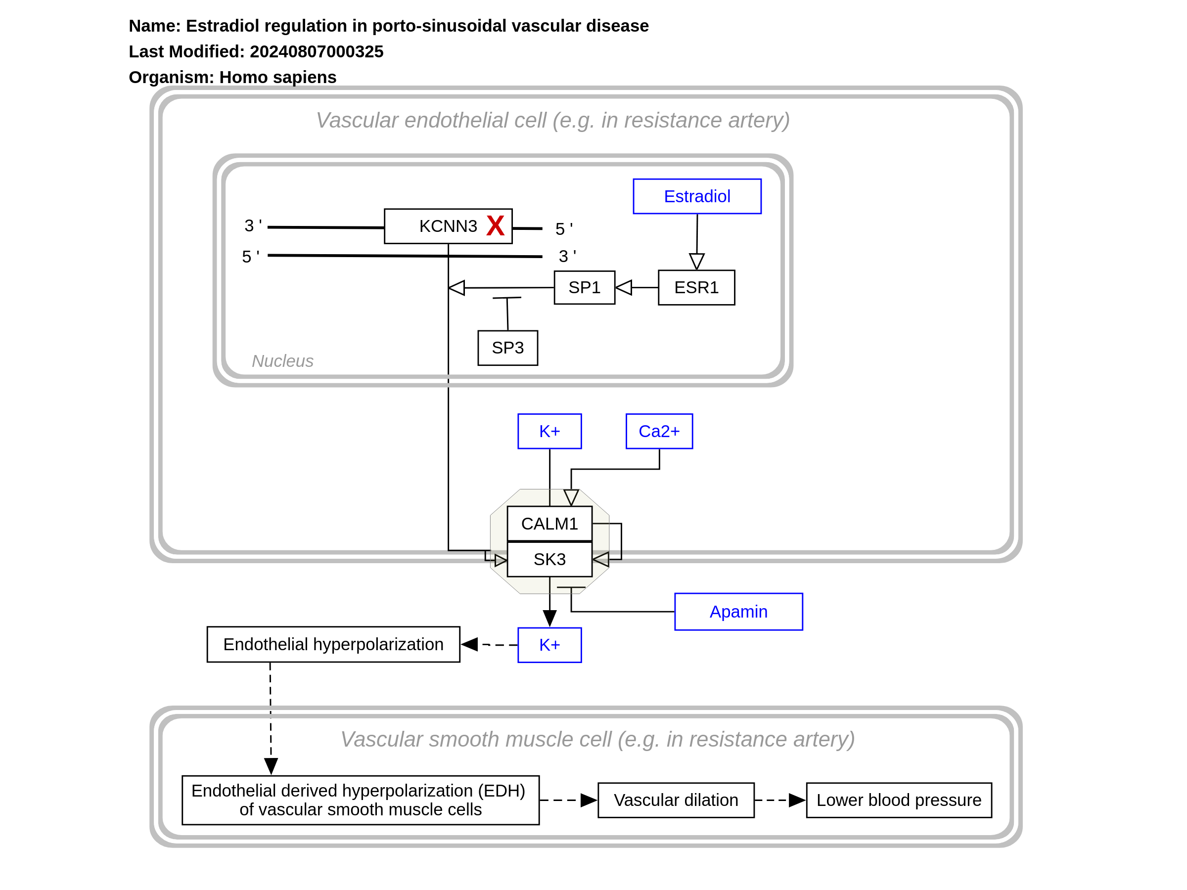
Porto-sinusoidal vascular disease (PSVD) is a rare disease (Schouten et al., 2015), affecting less than 1 in 2000 citizens (European standard) (Griffon et al., 2016). It is characterized by signs of presinusoidal portal hypertension without cirrhosis, where the cause of the hypertension is unknown (Lee et al., 2016; Schouten et al., 2011). Based on that what is known about the etiology of PSVD, its development can be categorized into five groups: immunological disorders, chronic infections, exposure to medications or toxins, prothrombic conditions, and genetic predisposition (Schouten et al., 2015). In practice, the disease has multiple contributing factors (Siramolpiwat et al., 2016). This pathway describes a mutation in the KCNN3 gene that is hypothesized to result in genetic predisposition to PSVD (Koot et al., 2016). PSVD has also been referred as idiopathic non-cirrhotic portal hypertension (INCPH), hepatoportal sclerosis, incomplete septal cirrhosis, obliterative portal venopathy, partial nodular transformation, non-cirrhotic portal fibrosis, nodular regenerative hyperplasia (NRH), and idiopathic portal hypertension (Schouten et al., 2015; Siramolpiwat et al., 2016; Besmond et al., 2017).
For a description of pathway objects, see the WikiPathways Legend.
Authors
ChristeldeVries , Friederike Ehrhart , Alex Pico , Egon Willighagen , Eric Weitz , Lars Willighagen , and Kristina HanspersActivity

Discuss this pathway
Check for ongoing discussions or start your own.
Cited In
Are you planning to include this pathway in your next publication? See How to Cite and add a link here to your paper once it's online.
Organisms
Homo sapiensCommunities
DiseasesAnnotations
Cell Type Ontology
endothelial cell vascular associated smooth muscle cellPathway Ontology
disease pathway hypertension pathwayDisease Ontology
portal hypertension vascular disease| Label | Type | Compact URI | Comment |
|---|---|---|---|
| Ca²⁺ | Metabolite | chebi:29108 | |
| Apamin | Metabolite | pubchem.compound:16133797 | The SK3 channel is apamin sensitive. The peptide toxin apamin, present in bee venom, blocks the SK3 channels, reducing hyperpolarization of the cells (Morikawa, 2010; Weisbrod, 2020) |
| Estradiol | Metabolite | chebi:16469 | |
| K⁺ | Metabolite | chebi:29103 | |
| K+ | Metabolite | chebi:29103 | |
| KCNN3 | GeneProduct | ensembl:ENSG00000143603 | A single nucleotide substitution (SNP) c.1348G>C in the KCNN3 gene was found in a father with INCPH and three of his children that were tested with Sanger- and whole-exome sequencing. These three children had developed signs of INCPH in the first ten years of their lives. A full sister of one of the girls had no complaints and did not participate in the examinations. Ultrasound and liver tests of the paternal grandparents showed no abnormalities of the liver. (Koot et al. 2015) KCNN genes are expressed in neurons, epithelium, endothelium of the vasculature, and several types of smooth muscle. The SK3 channel, which is a gene product of the KCNN3 gene, is involved in vascular tone- and blood pressure regulation. Calcium-induced activation of the SK3 channel will cause hyperpolarization of endothelial cells, resulting in hyperpolarization of the adjacent muscle cell, which is also known as an endothelium-derived hyperpolarizing factor (EDHF). This hyperpolarization of the muscle cells will then result in dilation in resistance arteries (Ledoux et al. 2006; Kohler et al. 2010). The level of SK3 channel expression in endothelial cells was found to be important for vascular tone and blood pressure in mice. (Taylor et al. 2003) Gene transcription: Two binding sites for the SP1 and SP3 transcription factors are present in the promotor region of the KCNN3 gene in mice. SP1 and SP3 compete to regulate the expression of the KCNN3 gene, influenced by the environment of the promotors. SP1 activates the expression of the gene, and SP3 inhibits the expression of the gene (Pierce et al., 2010; Xiong et al., 2020) . It was found that ERα stimulates the transcription KCNN3 through these transcription factors (Jacobson et al., 2003). |
| ESR1 | Protein | uniprot:P03372 | |
| SP1 | Protein | uniprot:P08047 | |
| SP3 | Protein | uniprot:Q02447 | |
| SK3 | Protein | uniprot:Q9UGI6 | Also referred to as: Small-conductance Ca2+- activated K+ (SK) channel Small conductance calcium-activated potassium channel protein 3 SK3 channel SKCa 3 KCa channel About: The SK3 channel is important for afterhyperpolarization following an action potential. One channel is made from four monomers which all contain six transmembrane segments, connected via a single pore loop. The N-termini and the C-termini are both located at the intracellular side of the membrane. A calmodulin molecule is located at the C-termini of the SK3 channel via a CaM-binding domain. Calmodulin will activate the SK3 channel upon binding of Ca²⁺. SK3 channels are not voltage-dependent. (Gu et al. 2018; Köhler et al. 1996; Weisbrod, 2020) Calcium-induced activation of the SK3 channel will cause hyperpolarization of endothelial cells, resulting in hyperpolarization of the adjacent muscle cell, which is also known as an endothelium-derived hyperpolarizing factor (EDHF). This hyperpolarization of the muscle cells will then result in dilation in resistance arteries (Ledoux et al. 2006; Kohler et al. 2010). The level of SK3 channel expression in endothelial cells was found to be important for vascular tone and blood pressure in mice. (Taylor et al. 2003) |
| CALM1 | Protein | uniprot:P0DP23 |
References
- Small-conductance, calcium-activated potassium channels from mammalian brain. Köhler M, Hirschberg B, Bond CT, Kinzie JM, Marrion NV, Maylie J, et al. Science. 1996 Sep 20;273(5282):1709–14. PubMed Europe PMC Scholia
- Determinants contributing to estrogen-regulated expression of SK3. Jacobson D, Pribnow D, Herson PS, Maylie J, Adelman JP. Biochem Biophys Res Commun. 2003 Apr 4;303(2):660–8. PubMed Europe PMC Scholia
- Altered expression of small-conductance Ca2+-activated K+ (SK3) channels modulates arterial tone and blood pressure. Taylor MS, Bonev AD, Gross TP, Eckman DM, Brayden JE, Bond CT, et al. Circ Res. 2003 Jul 25;93(2):124–31. PubMed Europe PMC Scholia
- Calcium-activated potassium channels and the regulation of vascular tone. Ledoux J, Werner ME, Brayden JE, Nelson MT. Physiology (Bethesda). 2006 Feb;21:69–78. PubMed Europe PMC Scholia
- Endothelial dysfunction and blood pressure alterations in K+-channel transgenic mice. Köhler R, Ruth P. Pflugers Arch. 2010 May;459(6):969–76. PubMed Europe PMC Scholia
- SK3 channel expression during pregnancy is regulated through estrogen and Sp factor-mediated transcriptional control of the KCNN3 gene. Pierce SL, England SK. Am J Physiol Endocrinol Metab. 2010 Oct;299(4):E640-6. PubMed Europe PMC Scholia
- Ethanol action on dopaminergic neurons in the ventral tegmental area: interaction with intrinsic ion channels and neurotransmitter inputs. Morikawa H, Morrisett RA. Int Rev Neurobiol. 2010;91:235–88. PubMed Europe PMC Scholia
- Idiopathic noncirrhotic portal hypertension. Schouten JNL, Garcia-Pagan JC, Valla DC, Janssen HLA. Hepatology. 2011 Sep 2;54(3):1071–81. PubMed Europe PMC Scholia
- Idiopathic non-cirrhotic portal hypertension: a review. Schouten JNL, Verheij J, Seijo S. Orphanet J Rare Dis. 2015 May 30;10:67. PubMed Europe PMC Scholia
- Idiopathic Noncirrhotic Portal Hypertension: An Appraisal. Lee H, Rehman AU, Fiel MI. J Pathol Transl Med. 2016 Jan;50(1):17–25. PubMed Europe PMC Scholia
- A de novo mutation in KCNN3 associated with autosomal dominant idiopathic non-cirrhotic portal hypertension. Koot BGP, Alders M, Verheij J, Beuers U, Cobben JM. J Hepatol. 2016 Apr;64(4):974–7. PubMed Europe PMC Scholia
- Searching for rare diseases in PubMed: a blind comparison of Orphanet expert query and query based on terminological knowledge. Griffon N, Schuers M, Dhombres F, Merabti T, Kerdelhué G, Rollin L, et al. BMC Med Inform Decis Mak. 2016 Aug 2;16:101. PubMed Europe PMC Scholia
- Mutations in the novel gene FOPV are associated with familial autosomal dominant and non-familial obliterative portal venopathy. Besmond C, Valla D, Hubert L, Poirier K, Grosse B, Guettier C, et al. Liver Int. 2018 Feb;38(2):358–64. PubMed Europe PMC Scholia
- Small-conductance Ca2+-activated K+ channels: insights into their roles in cardiovascular disease. Gu M, Zhu Y, Yin X, Zhang DM. Exp Mol Med. 2018 Apr 13;50(4):1–7. PubMed Europe PMC Scholia
- Small and Intermediate Calcium Activated Potassium Channels in the Heart: Role and Strategies in the Treatment of Cardiovascular Diseases. Weisbrod D. Front Physiol. 2020 Nov 23;11:590534. PubMed Europe PMC Scholia